1G9X

CHARACTERIZATION OF THE TWINNING STRUCTURE OF MJ1267, AN ATP-BINDING CASSETTE OF AN ABC TRANSPORTER


Experimental Data Snapshot

  • Method: X-RAY DIFFRACTION
  • Resolution: 2.60 Å
  • R-Value Free: 0.279 
  • R-Value Work: 0.153 
  • R-Value Observed: 0.153 

wwPDB Validation   3D Report Full Report


Ligand Structure Quality Assessment 


This is version 1.5 of the entry. See complete history


Literature

Structural characterization of an MJ1267 ATP-binding cassette crystal with a complex pattern of twinning caused by promiscuous fiber packing.

Yuan, Y.R.Martsinkevich, O.Hunt, J.F.

(2003) Acta Crystallogr D Biol Crystallogr 59: 225-238

  • DOI: https://doi.org/10.1107/s0907444902018954
  • Primary Citation of Related Structures:  
    1G9X

  • PubMed Abstract: 

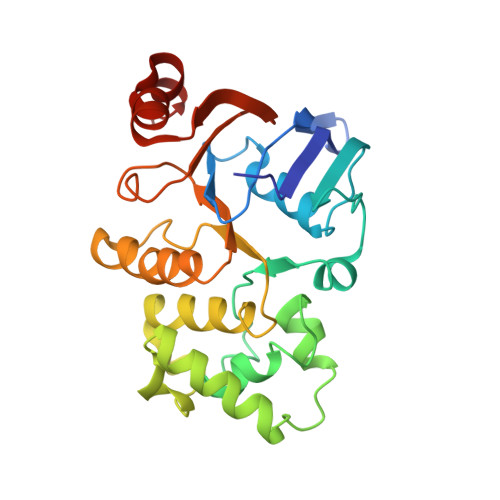
    ATP-binding cassettes represent the motor domains in ABC transporters, a superfamily of integral membrane-protein pumps that couple the hydrolysis of ATP to transmembrane solute translocation. A crystal of a Mg-ADP complex of the MJ1267 ATP-binding cassette was obtained that produced a diffraction pattern characterized by pathological streaking of the spots in the a* x b* plane. While the Laue symmetry of the diffraction pattern was P3;1m, the crystal was determined to be twinned based on intensity statistics, molecular-replacement analysis and difference Fourier analysis of an engineered single-site methylmercury derivative. The unit cell contains three similar 3(1) fibers, with two of them related by primarily translational non-crystallographic symmetry (NCS) and the third related to the first two by approximate twofold screw operations whose rotational components are very similar to the twinning operator. The promiscuous packing of these 3(1) fibers, which make both parallel and antiparallel interactions in the primary crystal lattice, can explain the twinning tendency based on the ability of the twin-related lattices to interact with one another while making only one slightly sub-optimal intermolecular contact per unit cell in the boundary region. The promiscuous fiber packing can also explain the streaking in the diffraction pattern based on the ability to form a variety of different lattices with similar inter-fiber packing interactions. The crystal structure was refined as a twin in space group P3(1) using the program CNS, yielding a free R factor of 28.9% at 2.6 A and a refined twin fraction of 0.50. The structure shows a rigid-body rotation of the ABC-transporter-specific alpha-helical subdomain (ABCalpha subdomain) in MJ1267 compared with the conformation observed for the same protein in a C2 crystal lattice; this observation suggests that the ABCalpha subdomain is flexibly attached to the F1-type ATP-binding core of the ATP-binding cassette when Mg-ADP is bound at the active site.


  • Organizational Affiliation

    Department of Biological Sciences, 702A Fairchild Center, MC2434, Columbia University, New York, NY 10027, USA.


Macromolecules
Find similar proteins by:  (by identity cutoff)  |  3D Structure
Entity ID: 1
MoleculeChains Sequence LengthOrganismDetailsImage
HIGH-AFFINITY BRANCHED-CHAIN AMINO ACID TRANSPORT ATP-BINDING PROTEIN
A, B, C
257Methanocaldococcus jannaschiiMutation(s): 1 
Gene Names: STRUCTURAL DNA
UniProt
Find proteins for Q58663 (Methanocaldococcus jannaschii (strain ATCC 43067 / DSM 2661 / JAL-1 / JCM 10045 / NBRC 100440))
Explore Q58663 
Go to UniProtKB:  Q58663
Entity Groups  
Sequence Clusters30% Identity50% Identity70% Identity90% Identity95% Identity100% Identity
UniProt GroupQ58663
Sequence Annotations
Expand
  • Reference Sequence
Small Molecules
Ligands 3 Unique
IDChains Name / Formula / InChI Key2D Diagram3D Interactions
ADP
Query on ADP

Download Ideal Coordinates CCD File 
F [auth A],
I [auth B],
L [auth C]
ADENOSINE-5'-DIPHOSPHATE
C10 H15 N5 O10 P2
XTWYTFMLZFPYCI-KQYNXXCUSA-N
MMC
Query on MMC

Download Ideal Coordinates CCD File 
E [auth A],
H [auth B],
K [auth C]
METHYL MERCURY ION
C H3 Hg
DBUXSCUEGJMZAE-UHFFFAOYSA-N
MG
Query on MG

Download Ideal Coordinates CCD File 
D [auth A],
G [auth B],
J [auth C]
MAGNESIUM ION
Mg
JLVVSXFLKOJNIY-UHFFFAOYSA-N
Experimental Data & Validation

Experimental Data

  • Method: X-RAY DIFFRACTION
  • Resolution: 2.60 Å
  • R-Value Free: 0.279 
  • R-Value Work: 0.153 
  • R-Value Observed: 0.153 
  • Space Group: P 31
Unit Cell:
Length ( Å )Angle ( ˚ )
a = 117.31α = 90
b = 117.31β = 90
c = 42.19γ = 120
Software Package:
Software NamePurpose
DENZOdata reduction
SCALEPACKdata scaling
AMoREphasing
CNSrefinement

Structure Validation

View Full Validation Report


Ligand Structure Quality Assessment 


Entry History 

Deposition Data

Revision History  (Full details and data files)

  • Version 1.0: 2003-02-11
    Type: Initial release
  • Version 1.1: 2008-04-01
    Changes: Version format compliance
  • Version 1.2: 2011-07-13
    Changes: Version format compliance
  • Version 1.3: 2021-10-27
    Changes: Database references, Derived calculations
  • Version 1.4: 2024-02-07
    Changes: Data collection
  • Version 1.5: 2024-04-03
    Changes: Refinement description