1G4A

CRYSTAL STRUCTURES OF THE HSLVU PEPTIDASE-ATPASE COMPLEX REVEAL AN ATP-DEPENDENT PROTEOLYSIS MECHANISM


Experimental Data Snapshot

  • Method: X-RAY DIFFRACTION
  • Resolution: 3.00 Å
  • R-Value Free: 0.294 
  • R-Value Work: 0.257 
  • R-Value Observed: 0.257 

wwPDB Validation   3D Report Full Report


Ligand Structure Quality Assessment 


This is version 1.3 of the entry. See complete history

Re-refinement Note

A newer entry is available that reflects an alternative modeling of the original data: 5JI3


Literature

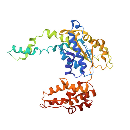
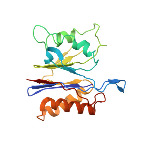
Crystal structures of the HslVU peptidase-ATPase complex reveal an ATP-dependent proteolysis mechanism.

Wang, J.Song, J.J.Franklin, M.C.Kamtekar, S.Im, Y.J.Rho, S.H.Seong, I.S.Lee, C.S.Chung, C.H.Eom, S.H.

(2001) Structure 9: 177-184

  • DOI: https://doi.org/10.1016/s0969-2126(01)00570-6
  • Primary Citation of Related Structures:  
    1G4A, 1G4B

  • PubMed Abstract: 

    The bacterial heat shock locus HslU ATPase and HslV peptidase together form an ATP-dependent HslVU protease. Bacterial HslVU is a homolog of the eukaryotic 26S proteasome. Crystallographic studies of HslVU should provide an understanding of ATP-dependent protein unfolding, translocation, and proteolysis by this and other ATP-dependent proteases.


  • Organizational Affiliation

    Department of Molecular Biophysics, Biochemistry, 266 Whitney Avenue, Yale University, 06520, New Haven, CT, USA. wang@mail.csb.yale.edu


Macromolecules
Find similar proteins by:  (by identity cutoff)  |  3D Structure
Entity ID: 1
MoleculeChains Sequence LengthOrganismDetailsImage
ATP-DEPENDENT HSL PROTEASE ATP-BINDING SUBUNIT HSLUA [auth E],
B [auth F]
443Escherichia coliMutation(s): 0 
UniProt
Find proteins for P0A6H5 (Escherichia coli (strain K12))
Explore P0A6H5 
Go to UniProtKB:  P0A6H5
Entity Groups  
Sequence Clusters30% Identity50% Identity70% Identity90% Identity95% Identity100% Identity
UniProt GroupP0A6H5
Sequence Annotations
Expand
  • Reference Sequence
Find similar proteins by:  (by identity cutoff)  |  3D Structure
Entity ID: 2
MoleculeChains Sequence LengthOrganismDetailsImage
ATP-DEPENDENT PROTEASE HSLVC [auth B],
D [auth A],
E [auth D],
F [auth C]
175Escherichia coliMutation(s): 0 
EC: 3.4.99
UniProt
Find proteins for P0A7B8 (Escherichia coli (strain K12))
Explore P0A7B8 
Go to UniProtKB:  P0A7B8
Entity Groups  
Sequence Clusters30% Identity50% Identity70% Identity90% Identity95% Identity100% Identity
UniProt GroupP0A7B8
Sequence Annotations
Expand
  • Reference Sequence
Small Molecules
Ligands 1 Unique
IDChains Name / Formula / InChI Key2D Diagram3D Interactions
DAT
Query on DAT

Download Ideal Coordinates CCD File 
G [auth E],
H [auth F]
2'-DEOXYADENOSINE-5'-DIPHOSPHATE
C10 H15 N5 O9 P2
DAEAPNUQQAICNR-RRKCRQDMSA-N
Experimental Data & Validation

Experimental Data

  • Method: X-RAY DIFFRACTION
  • Resolution: 3.00 Å
  • R-Value Free: 0.294 
  • R-Value Work: 0.257 
  • R-Value Observed: 0.257 
  • Space Group: P 3 2 1
Unit Cell:
Length ( Å )Angle ( ˚ )
a = 169.995α = 90
b = 169.995β = 90
c = 161.317γ = 120
Software Package:
Software NamePurpose
X-PLORmodel building
CNSrefinement
DENZOdata reduction
SCALEPACKdata scaling
X-PLORphasing

Structure Validation

View Full Validation Report


Ligand Structure Quality Assessment 


Entry History 

Deposition Data

Revision History  (Full details and data files)

  • Version 1.0: 2001-02-21
    Type: Initial release
  • Version 1.1: 2008-04-27
    Changes: Version format compliance
  • Version 1.2: 2011-07-13
    Changes: Version format compliance
  • Version 1.3: 2024-02-07
    Changes: Data collection, Database references, Derived calculations