1FW2

OUTER MEMBRANE PHOSPHOLIPASE A FROM ESCHERICHIA COLI


Experimental Data Snapshot

  • Method: X-RAY DIFFRACTION
  • Resolution: 2.60 Å
  • R-Value Free: 0.298 
  • R-Value Work: 0.222 

wwPDB Validation   3D Report Full Report


This is version 1.3 of the entry. See complete history


Literature

Structural investigations of calcium binding and its role in activity and activation of outer membrane phospholipase A from Escherichia coli.

Snijder, H.J.Kingma, R.L.Kalk, K.H.Dekker, N.Egmond, M.R.Dijkstra, B.W.

(2001) J Mol Biol 309: 477-489

  • DOI: https://doi.org/10.1006/jmbi.2001.4675
  • Primary Citation of Related Structures:  
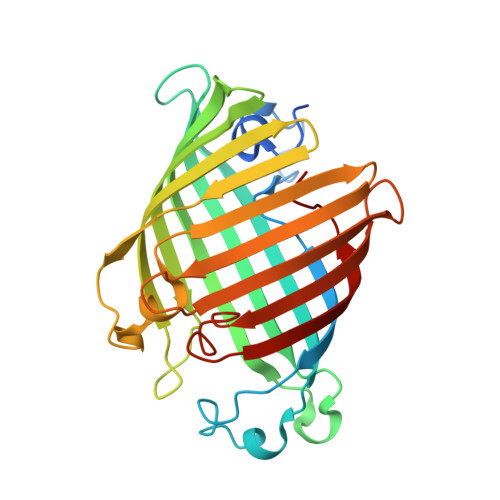
    1FW2, 1FW3

  • PubMed Abstract: 

    Outer membrane phospholipase A (OMPLA) is an integral membrane enzyme that catalyses the hydrolysis of phospholipids. Enzymatic activity is regulated by reversible dimerisation and calcium-binding. We have investigated the role of calcium by X-ray crystallography. In monomeric OMPLA, one calcium ion binds between two external loops (L3L4 site) at 10 A from the active site. After dimerisation, a new calcium-binding site (catalytic site) is formed at the dimer interface in the active site of each molecule at 6 A from the L3L4 calcium site. The close spacing and the difference in calcium affinity of both sites suggests that the L3L4 site may function as a storage site for a calcium ion, which relocates to the catalytic site upon dimerisation. A sequence alignment demonstrates conservation of the catalytic calcium site but evolutionary variation of the L3L4 site. The residues in the dimer interface are conserved as well, suggesting that all outer membrane phospholipases require dimerisation and calcium in the catalytic site for activity. For this family of phospholipases, we have characterised a consensus sequence motif (YTQ-X(n)-G-X(2)-H-X-SNG) that contains conserved residues involved in dimerisation and catalysis.


  • Organizational Affiliation

    Laboratory of Biophysical Chemistry, BIOSON Research Institute, University of Groningen, Nijenborgh 4 9747 AG, Groningen, The Netherlands. bauke@chem.rug.nl


Macromolecules
Find similar proteins by:  (by identity cutoff)  |  3D Structure
Entity ID: 1
MoleculeChains Sequence LengthOrganismDetailsImage
OUTER MEMBRANE PHOSPHOLIPASE A275Escherichia coliMutation(s): 0 
EC: 3.1.1.32
Membrane Entity: Yes 
UniProt
Find proteins for P0A921 (Escherichia coli (strain K12))
Explore P0A921 
Go to UniProtKB:  P0A921
Entity Groups  
Sequence Clusters30% Identity50% Identity70% Identity90% Identity95% Identity100% Identity
UniProt GroupP0A921
Sequence Annotations
Expand
  • Reference Sequence
Small Molecules
Ligands 1 Unique
IDChains Name / Formula / InChI Key2D Diagram3D Interactions
CA
Query on CA

Download Ideal Coordinates CCD File 
B [auth A]CALCIUM ION
Ca
BHPQYMZQTOCNFJ-UHFFFAOYSA-N
Experimental Data & Validation

Experimental Data

  • Method: X-RAY DIFFRACTION
  • Resolution: 2.60 Å
  • R-Value Free: 0.298 
  • R-Value Work: 0.222 
  • Space Group: P 31 2 1
Unit Cell:
Length ( Å )Angle ( ˚ )
a = 77.532α = 90
b = 77.532β = 90
c = 100.525γ = 120
Software Package:
Software NamePurpose
X-PLORmodel building
X-PLORrefinement
DENZOdata reduction
SCALEPACKdata scaling
X-PLORphasing

Structure Validation

View Full Validation Report


Entry History 

Deposition Data

Revision History  (Full details and data files)

  • Version 1.0: 2001-06-01
    Type: Initial release
  • Version 1.1: 2008-04-27
    Changes: Version format compliance
  • Version 1.2: 2011-07-13
    Changes: Version format compliance
  • Version 1.3: 2022-12-21
    Changes: Database references, Derived calculations