1EYW

THREE-DIMENSIONAL STRUCTURE OF THE ZINC-CONTAINING PHOSPHOTRIESTERASE WITH BOUND SUBSTRATE ANALOG TRIETHYLPHOSPHATE


Experimental Data Snapshot

  • Method: X-RAY DIFFRACTION
  • Resolution: 1.90 Å
  • R-Value Observed: 0.148 

wwPDB Validation   3D Report Full Report


This is version 1.3 of the entry. See complete history


Literature

The binding of substrate analogs to phosphotriesterase.

Benning, M.M.Hong, S.B.Raushel, F.M.Holden, H.M.

(2000) J Biol Chem 275: 30556-30560

  • DOI: https://doi.org/10.1074/jbc.M003852200
  • Primary Citation of Related Structures:  
    1EYW, 1EZ2

  • PubMed Abstract: 

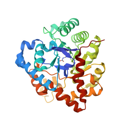
    Phosphotriesterase (PTE) from Pseudomonas diminuta catalyzes the detoxification of organophosphates such as the widely utilized insecticide paraoxon and the chemical warfare agent sarin. The three-dimensional structure of the enzyme is known from high resolution x-ray crystallographic analyses. Each subunit of the homodimer folds into a so-called TIM barrel, with eight strands of parallel beta-sheet. The two zinc ions required for activity are positioned at the C-terminal portion of the beta-barrel. Here, we describe the three-dimensional structure of PTE complexed with the inhibitor diisopropyl methyl phosphonate, which serves as a mimic for sarin. Additionally, the structure of the enzyme complexed with triethyl phosphate is also presented. In the case of the PTE-diisopropyl methyl phosphonate complex, the phosphoryl oxygen of the inhibitor coordinates to the more solvent-exposed zinc ion (2.5 A), thereby lending support to the presumed catalytic mechanism involving metal coordination of the substrate. In the PTE-triethyl phosphate complex, the phosphoryl oxygen of the inhibitor is positioned at 3.4 A from the more solvent-exposed zinc ion. The two structures described in this report provide additional molecular understanding for the ability of this remarkable enzyme to hydrolyze such a wide range of organophosphorus substrates.


  • Organizational Affiliation

    Department of Biochemistry, University of Wisconsin, Madison, USA.


Macromolecules
Find similar proteins by:  (by identity cutoff)  |  3D Structure
Entity ID: 1
MoleculeChains Sequence LengthOrganismDetailsImage
PHOSPHOTRIESTERASE331Brevundimonas diminutaMutation(s): 1 
EC: 3.1.8.1
UniProt
Find proteins for P0A434 (Brevundimonas diminuta)
Explore P0A434 
Go to UniProtKB:  P0A434
Entity Groups  
Sequence Clusters30% Identity50% Identity70% Identity90% Identity95% Identity100% Identity
UniProt GroupP0A434
Sequence Annotations
Expand
  • Reference Sequence
Small Molecules
Ligands 3 Unique
IDChains Name / Formula / InChI Key2D Diagram3D Interactions
TEN
Query on TEN

Download Ideal Coordinates CCD File 
D [auth A]TRIETHYL PHOSPHATE
C6 H15 O4 P
DQWPFSLDHJDLRL-UHFFFAOYSA-N
PEL
Query on PEL

Download Ideal Coordinates CCD File 
E [auth A]2-PHENYL-ETHANOL
C8 H10 O
WRMNZCZEMHIOCP-UHFFFAOYSA-N
ZN
Query on ZN

Download Ideal Coordinates CCD File 
B [auth A],
C [auth A]
ZINC ION
Zn
PTFCDOFLOPIGGS-UHFFFAOYSA-N
Modified Residues  1 Unique
IDChains TypeFormula2D DiagramParent
KCX
Query on KCX
A
L-PEPTIDE LINKINGC7 H14 N2 O4LYS
Experimental Data & Validation

Experimental Data

  • Method: X-RAY DIFFRACTION
  • Resolution: 1.90 Å
  • R-Value Observed: 0.148 
  • Space Group: C 2 2 21
Unit Cell:
Length ( Å )Angle ( ˚ )
a = 128.62α = 90
b = 92.33β = 90
c = 69.85γ = 90
Software Package:
Software NamePurpose
TNTrefinement
FRAMBOdata collection
XDSdata scaling
TNTphasing

Structure Validation

View Full Validation Report


Entry History 

Deposition Data

Revision History  (Full details and data files)

  • Version 1.0: 2000-12-20
    Type: Initial release
  • Version 1.1: 2008-04-27
    Changes: Version format compliance
  • Version 1.2: 2011-07-13
    Changes: Version format compliance
  • Version 1.3: 2017-10-04
    Changes: Refinement description