Crystal structures of substrate free and complex forms of reactivated BphC, an extradiol type ring-cleavage dioxygenase.
Uragami, Y., Senda, T., Sugimoto, K., Sato, N., Nagarajan, V., Masai, E., Fukuda, M., Mitsu, Y.(2001) J Inorg Biochem 83: 269-279
- PubMed: 11293547
- DOI: https://doi.org/10.1016/s0162-0134(00)00172-0
- Primary Citation of Related Structures:
1EIL, 1EIQ, 1EIR - PubMed Abstract:
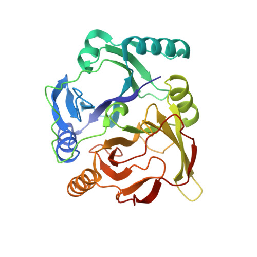
BphC derived from Pseudomonas sp. strain KKS102, an extradiol type catecholic dioxygenase, is a non-heam iron-containing enzyme, playing an important role in the degradation of biphenyl/PCB (Poly Chlorinated Biphenyls) in the microbe. Although we had earlier solved the crystal structure of KKS102 BphC, it was the inactive form with Fe(III) in the active site. In order to determine the active form structure, BphC was re-activated by anaerobic incubation with Fe(II) and ascorbate, and crystallized anaerobically. The crystal structures of activated BphC and its substrate complex (E x S complex) were determined at 2.0 A resolution under cryogenic condition. In addition, crystal structures of unactivated BphC in substrate free and complex forms were also re-determined. Comparison of activated and unactivated E x S complexes reveals that the orientation of the bound substrate in the active site is significantly different between the two. The structural comparison of the substrate free and complex forms of activated BphC show certain small conformational shifts around the active site upon substrate binding. As a result of the conformational shifts, His194, which has been suggested as the catalytic base, takes part in a weak hydrogen bond with hydroxyl group of the substrate.
Organizational Affiliation:
Department of BioEngineering, Nagaoka University of Technology, Niigata, Japan.