1E9O

Crystal structure of bovine SOD - 1 of 3


Experimental Data Snapshot

  • Method: X-RAY DIFFRACTION
  • Resolution: 1.85 Å
  • R-Value Free: 0.230 
  • R-Value Work: 0.199 
  • R-Value Observed: 0.199 

wwPDB Validation   3D Report Full Report


This is version 1.2 of the entry. See complete history


Literature

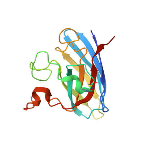
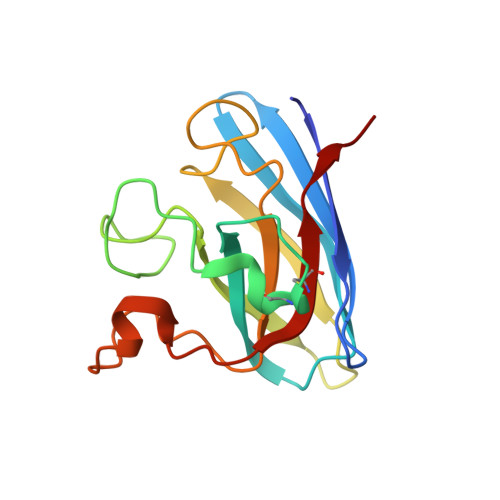
Conformational Variability of the Cu Site in One Subunit of Bovine Cuzn Superoxide Dismutase: The Importance of Mobility in the Glu119-Leu142 Loop Region for Catalytic Function

Hough, M.A.Strange, R.W.Hasnain, S.S.

(2000) J Mol Biol 304: 231

  • DOI: https://doi.org/10.1006/jmbi.2000.4186
  • Primary Citation of Related Structures:  
    1E9O, 1E9P, 1E9Q

  • PubMed Abstract: 

    The structure of the catalytic site in one subunit of bovine CuZn superoxide dismutase is shown to be highly variable. A series of crystal structures at approximately 1.7 A have been determined using data collected from different crystals. Several conformations are observed for the copper site from one of the subunits. These conformations lie between those expected for the Cu(II) and Cu(I) forms of the enzyme and may represent a slow positional rearrangement of the Cu site during the crystallisation process due to the presence of a trace reductant in the mother liquor. These states perhaps indicate some functionally relevant structural steps that ultimately result in the breakage of the imidazolate bridge between the two metal sites. This behaviour is not observed for the second subunit of the dimeric enzyme, which remains in the five-coordinate, distorted square planar geometry in all cases. We suggest that this asymmetric behaviour may be caused by the lack of mobility for the Glu119-Leu142 loop region in the second subunit caused by crystal contacts. This region forms one wall of the active-site cavity, and its mobility has been suggested, via molecular dynamics studies, to be important for the catalytic mechanism.


  • Organizational Affiliation

    CLRC Daresbury Laboratory, Daresbury, Warrington, WA4 4AD, UK.


Macromolecules
Find similar proteins by:  (by identity cutoff)  |  3D Structure
Entity ID: 1
MoleculeChains Sequence LengthOrganismDetailsImage
SUPEROXIDE DISMUTASE152Bos taurusMutation(s): 0 
EC: 1.15.1.1
UniProt
Find proteins for P00442 (Bos taurus)
Explore P00442 
Go to UniProtKB:  P00442
Entity Groups  
Sequence Clusters30% Identity50% Identity70% Identity90% Identity95% Identity100% Identity
UniProt GroupP00442
Sequence Annotations
Expand
  • Reference Sequence
Find similar proteins by:  (by identity cutoff)  |  3D Structure
Entity ID: 2
MoleculeChains Sequence LengthOrganismDetailsImage
SUPEROXIDE DISMUTASE152Bos taurusMutation(s): 0 
EC: 1.15.1.1
UniProt
Find proteins for P00442 (Bos taurus)
Explore P00442 
Go to UniProtKB:  P00442
Entity Groups  
Sequence Clusters30% Identity50% Identity70% Identity90% Identity95% Identity100% Identity
UniProt GroupP00442
Sequence Annotations
Expand
  • Reference Sequence
Experimental Data & Validation

Experimental Data

  • Method: X-RAY DIFFRACTION
  • Resolution: 1.85 Å
  • R-Value Free: 0.230 
  • R-Value Work: 0.199 
  • R-Value Observed: 0.199 
  • Space Group: P 21 21 21
Unit Cell:
Length ( Å )Angle ( ˚ )
a = 47.5α = 90
b = 51β = 90
c = 147.8γ = 90
Software Package:
Software NamePurpose
REFMACrefinement
DENZOdata reduction
SCALEPACKdata scaling
AMoREphasing

Structure Validation

View Full Validation Report


Entry History 

Deposition Data

Revision History  (Full details and data files)

  • Version 1.0: 2000-12-03
    Type: Initial release
  • Version 1.1: 2015-09-16
    Changes: Data collection, Derived calculations, Non-polymer description, Other, Refinement description, Source and taxonomy, Version format compliance
  • Version 1.2: 2023-12-13
    Changes: Data collection, Database references, Derived calculations, Refinement description