1DU3

Crystal structure of TRAIL-SDR5


Experimental Data Snapshot

  • Method: X-RAY DIFFRACTION
  • Resolution: 2.20 Å
  • R-Value Free: 0.291 
  • R-Value Work: 0.291 
  • R-Value Observed: 0.212 

wwPDB Validation   3D Report Full Report


This is version 1.3 of the entry. See complete history


Literature

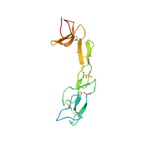
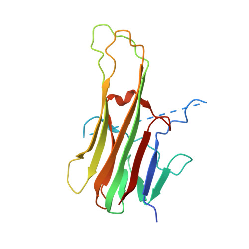
Crystal structure of TRAIL-DR5 complex identifies a critical role of the unique frame insertion in conferring recognition specificity

Cha, S.-S.Sung, B.-J.Kim, Y.A.Song, Y.L.Kim, H.J.Kim, S.Lee, M.S.Oh, B.-H.

(2000) J Biol Chem 275: 31171-31177

  • DOI: https://doi.org/10.1074/jbc.M004414200
  • Primary Citation of Related Structures:  
    1DU3

  • PubMed Abstract: 

    TRAIL is a cytokine that induces apoptosis in a wide variety of tumor cells but rarely in normal cells. It contains an extraordinarily elongated loop because of an unique insertion of 12-16 amino acids compared with the other members of tumor necrosis factor family. Biological implication of the frame insertion has not been clarified. We have determined the crystal structure of TRAIL in a complex with the extracellular domain of death receptor DR5 at 2.2 A resolution. The structure reveals extensive contacts between the elongated loop and DR5 in an interaction mode that would not be allowed without the frame insertion. These interactions are missing in the structures of the complex determined by others recently. This observation, along with structure-inspired deletion analysis, identifies the critical role of the frame insertion as a molecular strategy conferring specificity upon the recognition of cognate receptors. The structure also suggests that a built-in flexibility of the tumor necrosis factor receptor family members is likely to play a general and important role in the binding and recognition of tumor necrosis factor family members.


  • Organizational Affiliation

    Department of Life Science, and Division of Molecular and Life Science, Pohang University of Science and Technology, Pohang, Kyungbuk, 790-784, Korea.


Macromolecules
Find similar proteins by:  (by identity cutoff)  |  3D Structure
Entity ID: 1
MoleculeChains Sequence LengthOrganismDetailsImage
DEATH RECEPTOR 5
A, B, C, G, H
A, B, C, G, H, I
130Homo sapiensMutation(s): 0 
UniProt & NIH Common Fund Data Resources
Find proteins for O14763 (Homo sapiens)
Explore O14763 
Go to UniProtKB:  O14763
PHAROS:  O14763
GTEx:  ENSG00000120889 
Entity Groups  
Sequence Clusters30% Identity50% Identity70% Identity90% Identity95% Identity100% Identity
UniProt GroupO14763
Sequence Annotations
Expand
  • Reference Sequence
Find similar proteins by:  (by identity cutoff)  |  3D Structure
Entity ID: 2
MoleculeChains Sequence LengthOrganismDetailsImage
TNF-RELATED APOPTOSIS INDUCING LIGAND
D, E, F, J, K
D, E, F, J, K, L
168Homo sapiensMutation(s): 0 
UniProt & NIH Common Fund Data Resources
Find proteins for P50591 (Homo sapiens)
Explore P50591 
Go to UniProtKB:  P50591
PHAROS:  P50591
GTEx:  ENSG00000121858 
Entity Groups  
Sequence Clusters30% Identity50% Identity70% Identity90% Identity95% Identity100% Identity
UniProt GroupP50591
Sequence Annotations
Expand
  • Reference Sequence
Small Molecules
Ligands 1 Unique
IDChains Name / Formula / InChI Key2D Diagram3D Interactions
ZN
Query on ZN

Download Ideal Coordinates CCD File 
M [auth E],
N [auth K]
ZINC ION
Zn
PTFCDOFLOPIGGS-UHFFFAOYSA-N
Experimental Data & Validation

Experimental Data

  • Method: X-RAY DIFFRACTION
  • Resolution: 2.20 Å
  • R-Value Free: 0.291 
  • R-Value Work: 0.291 
  • R-Value Observed: 0.212 
  • Space Group: P 1 21 1
Unit Cell:
Length ( Å )Angle ( ˚ )
a = 68.63α = 90
b = 124.81β = 104.489
c = 128.37γ = 90
Software Package:
Software NamePurpose
DENZOdata reduction
SCALEPACKdata scaling
AMoREphasing
X-PLORrefinement

Structure Validation

View Full Validation Report


Entry History 

Deposition Data

Revision History  (Full details and data files)

  • Version 1.0: 2000-09-27
    Type: Initial release
  • Version 1.1: 2008-04-27
    Changes: Version format compliance
  • Version 1.2: 2011-07-13
    Changes: Version format compliance
  • Version 1.3: 2018-01-31
    Changes: Experimental preparation