1DPP

DIPEPTIDE BINDING PROTEIN COMPLEX WITH GLYCYL-L-LEUCINE


Experimental Data Snapshot

  • Method: X-RAY DIFFRACTION
  • Resolution: 3.20 Å
  • R-Value Free: 0.240 
  • R-Value Work: 0.223 
  • R-Value Observed: 0.223 

wwPDB Validation   3D Report Full Report


This is version 1.2 of the entry. See complete history


Literature

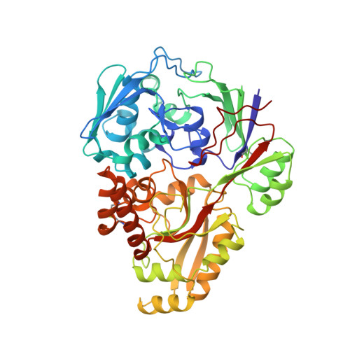
Crystal structure of the dipeptide binding protein from Escherichia coli involved in active transport and chemotaxis.

Dunten, P.Mowbray, S.L.

(1995) Protein Sci 4: 2327-2334

  • DOI: https://doi.org/10.1002/pro.5560041110
  • Primary Citation of Related Structures:  
    1DPP

  • PubMed Abstract: 

    The Escherichia coli periplasmic dipeptide binding protein functions in both peptide transport and taxis toward peptides. The structure of the dipeptide binding protein in complex with Gly-Leu (glycyl-L-leucine) has been determined at 3.2 A resolution. The binding site for dipeptides is designed to recognize the ligand's backbone while providing space to accommodate a variety of side chains. Some repositioning of protein side chains lining the binding site must occur when the dipeptide's second residue is larger than leucine. The protein's fold is very similar to that of the Salmonella typhimurium oligopeptide binding protein, and a comparison of the structures reveals the structural basis for the dipeptide binding protein's preference for shorter peptides.


  • Organizational Affiliation

    Department of Molecular Biology, Swedish University of Agricultural Sciences, Uppsala, Sweden. dunten@xray.bmc.uu.se


Macromolecules
Find similar proteins by:  (by identity cutoff)  |  3D Structure
Entity ID: 1
MoleculeChains Sequence LengthOrganismDetailsImage
DIPEPTIDE BINDING PROTEINA,
B [auth C],
C [auth E],
D [auth G]
507Escherichia coliMutation(s): 0 
UniProt
Find proteins for P23847 (Escherichia coli (strain K12))
Explore P23847 
Go to UniProtKB:  P23847
Entity Groups  
Sequence Clusters30% Identity50% Identity70% Identity90% Identity95% Identity100% Identity
UniProt GroupP23847
Sequence Annotations
Expand
  • Reference Sequence
Experimental Data & Validation

Experimental Data

  • Method: X-RAY DIFFRACTION
  • Resolution: 3.20 Å
  • R-Value Free: 0.240 
  • R-Value Work: 0.223 
  • R-Value Observed: 0.223 
  • Space Group: P 61
Unit Cell:
Length ( Å )Angle ( ˚ )
a = 182.57α = 90
b = 182.57β = 90
c = 211.88γ = 120
Software Package:
Software NamePurpose
DENZOdata reduction
X-PLORmodel building
X-PLORrefinement
X-PLORphasing

Structure Validation

View Full Validation Report


Entry History 

Deposition Data

Revision History  (Full details and data files)

  • Version 1.0: 1995-12-07
    Type: Initial release
  • Version 1.1: 2008-03-24
    Changes: Version format compliance
  • Version 1.2: 2011-07-13
    Changes: Version format compliance