1DMS

STRUCTURE OF DMSO REDUCTASE


Experimental Data Snapshot

  • Method: X-RAY DIFFRACTION
  • Resolution: 1.88 Å
  • R-Value Free: 0.203 
  • R-Value Work: 0.169 
  • R-Value Observed: 0.169 

wwPDB Validation   3D Report Full Report


This is version 2.1 of the entry. See complete history


Literature

Crystal structure of dimethyl sulfoxide reductase from Rhodobacter capsulatus at 1.88 A resolution.

Schneider, F.Lowe, J.Huber, R.Schindelin, H.Kisker, C.Knablein, J.

(1996) J Mol Biol 263: 53-69

  • DOI: https://doi.org/10.1006/jmbi.1996.0555
  • Primary Citation of Related Structures:  
    1DMS

  • PubMed Abstract: 

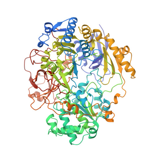
    The periplasmic dimethyl sulfoxide reductase (DMSOR) from the photosynthetic purple bacterium Rhodobacter capsulatus functions as the terminal electron acceptor in its respiratory chain. The enzyme catalyzes the reduction of highly oxidized substrates like dimethyl sulfoxide to dimethyl sulfide. At a molybdenum redox center, two single electrons are transferred from cytochrome C556 to the substrate dimethyl sulfoxide, generating dimethyl sulfide and (with two protons) water. The enzyme was purified and crystallized in space group P4(1)2(1)2 with unit cell dimensions of a = b = 80.7 A and c = 229.2 A. The crystals diffract beyond 1.8 A with synchrotron radiation. The three-dimensional structure was solved by a combination of multiple isomorphous replacement and molecular replacement techniques. The atomic model was refined to an R-factor of 0.169 for 57,394 independent reflections. The spherical protein consists of four domains with a funnel-like cavity that leads to the freely accessible metal-ion redox center. The bis(molybdopterin guanine dinucleotide) molybdenum cofactor (1541 Da) of the single chain protein (85,033 Da) has the molybdenum ion bound to the cis-dithiolene group of only one molybdopterin guanine dinucleotide molecule. Three additional ligands, two oxo groups and the oxygen of a serine side-chain, are bound to the molybdenum ion. The second molybdopterin system is not part of the ligand sphere of the metal center with its sulfur atoms at distances of 3.5 A and 3.8 A away. It might be involved in electron shuttling from the protein surface to the molybdenum center.


  • Organizational Affiliation

    Max-Planck-Institut für Biochemie, Martinsried, FRG.


Macromolecules
Find similar proteins by:  (by identity cutoff)  |  3D Structure
Entity ID: 1
MoleculeChains Sequence LengthOrganismDetailsImage
DMSO REDUCTASE781Rhodobacter capsulatusMutation(s): 0 
UniProt
Find proteins for Q52675 (Rhodobacter capsulatus)
Explore Q52675 
Go to UniProtKB:  Q52675
Entity Groups  
Sequence Clusters30% Identity50% Identity70% Identity90% Identity95% Identity100% Identity
UniProt GroupQ52675
Sequence Annotations
Expand
  • Reference Sequence
Small Molecules
Ligands 2 Unique
IDChains Name / Formula / InChI Key2D Diagram3D Interactions
PGD
Query on PGD

Download Ideal Coordinates CCD File 
B [auth A],
C [auth A]
2-AMINO-5,6-DIMERCAPTO-7-METHYL-3,7,8A,9-TETRAHYDRO-8-OXA-1,3,9,10-TETRAAZA-ANTHRACEN-4-ONE GUANOSINE DINUCLEOTIDE
C20 H24 N10 O13 P2 S2
SOFMTHSWCZNJTQ-ILXWUORBSA-N
2MO
Query on 2MO

Download Ideal Coordinates CCD File 
D [auth A]MOLYBDENUM (IV)OXIDE
Mo O2
QXYJCZRRLLQGCR-UHFFFAOYSA-N
Experimental Data & Validation

Experimental Data

  • Method: X-RAY DIFFRACTION
  • Resolution: 1.88 Å
  • R-Value Free: 0.203 
  • R-Value Work: 0.169 
  • R-Value Observed: 0.169 
  • Space Group: P 41 21 2
Unit Cell:
Length ( Å )Angle ( ˚ )
a = 80.7α = 90
b = 80.7β = 90
c = 229.2γ = 90
Software Package:
Software NamePurpose
X-PLORmodel building
X-PLORrefinement
MOSFLMdata reduction
X-PLORphasing

Structure Validation

View Full Validation Report


Entry History 

Deposition Data

Revision History  (Full details and data files)

  • Version 1.0: 1998-07-01
    Type: Initial release
  • Version 1.1: 2008-03-06
    Changes: Version format compliance
  • Version 1.2: 2011-07-13
    Changes: Version format compliance
  • Version 2.0: 2018-03-14
    Changes: Advisory, Atomic model, Data collection, Derived calculations, Non-polymer description, Other, Structure summary
  • Version 2.1: 2024-02-07
    Changes: Data collection, Database references, Derived calculations