Crystal structure of the ARF-GAP domain and ankyrin repeats of PYK2-associated protein beta.
Mandiyan, V., Andreev, J., Schlessinger, J., Hubbard, S.R.(1999) EMBO J 18: 6890-6898
- PubMed: 10601011
- DOI: https://doi.org/10.1093/emboj/18.24.6890
- Primary Citation of Related Structures:
1DCQ - PubMed Abstract:
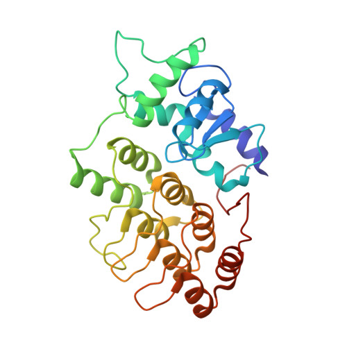
ADP ribosylation factors (ARFs), which are members of the Ras superfamily of GTP-binding proteins, are critical components of vesicular trafficking pathways in eukaryotes. Like Ras, ARFs are active in their GTP-bound form, and their duration of activity is controlled by GTPase-activating proteins (GAPs), which assist ARFs in hydrolyzing GTP to GDP. PAPbeta, a protein that binds to and is phosphorylated by the non-receptor tyrosine kinase PYK2, contains several modular signaling domains including a pleckstrin homology domain, an SH3 domain, ankyrin repeats and an ARF-GAP domain. Sequences of ARF-GAP domains show no recognizable similarity to those of other GAPs, and contain a characteristic Cys-X(2)-Cys-X(16-17)-Cys-X(2)-Cys motif. The crystal structure of the PAPbeta ARF-GAP domain and the C-terminal ankyrin repeats has been determined at 2.1 A resolution. The ARF-GAP domain comprises a central three-stranded beta-sheet flanked by five alpha-helices, with a Zn(2+) ion coordinated by the four cysteines of the cysteine-rich motif. Four ankyrin repeats are also present, the first two of which form an extensive interface with the ARF-GAP domain. An invariant arginine and several nearby hydrophobic residues are solvent exposed and are predicted to be the site of interaction with ARFs. Site-directed mutagenesis of these residues confirms their importance in ARF-GAP activity.
Organizational Affiliation:
Department of Pharmacology, Skirball Institute of Biomolecular Medicine, New York University Medical School, New York, NY 10016, USA.