1D3F

N-TERMINAL DOMAIN CORE METHIONINE MUTATION


Experimental Data Snapshot

  • Method: X-RAY DIFFRACTION
  • Resolution: 2.05 Å
  • R-Value Work: 0.159 
  • R-Value Observed: 0.159 

wwPDB Validation   3D Report Full Report


Ligand Structure Quality Assessment 


This is version 1.5 of the entry. See complete history


Literature

Use of differentially substituted selenomethionine proteins in X-ray structure determination.

Gassner, N.C.Matthews, B.W.

(1999) Acta Crystallogr D Biol Crystallogr 55: 1967-1970

  • DOI: https://doi.org/10.1107/s0907444999013347
  • Primary Citation of Related Structures:  
    1CX7, 1D2W, 1D2Y, 1D3F, 1D3J, 1D3M, 1D3N

  • PubMed Abstract: 

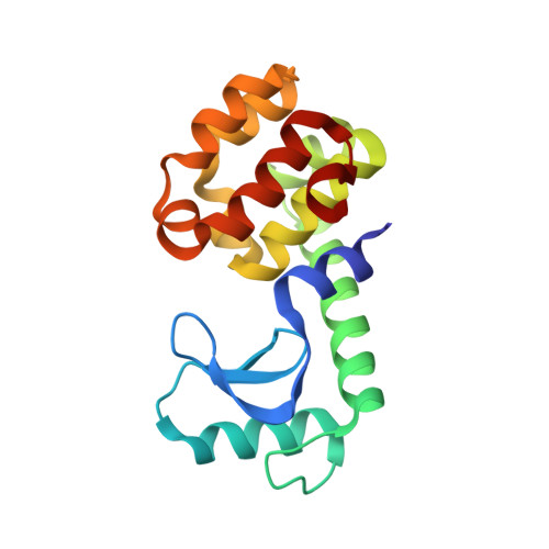
    Using heavily methionine-substituted T4 lysozyme as an example, it is shown how the addition or deletion of a small number of methionines can simplify the location of selenium sites for use in MAD phasing. By comparing the X-ray data for a large number of singly substituted lysozymes, it is shown that the optimal amino acid to be substituted by methionine is leucine, followed, in order of preference, by phenylalanine, isoleucine and valine. The identification of leucine as the first choice agrees with the ranking suggested by the Dayhoff mutation probability, i.e. by the frequency of amino-acid substitutions in the sequences of related proteins. The ranking of the second and subsequent choices, however, differ significantly.


  • Organizational Affiliation

    Institute of Molecular Biology, Howard Hughes Medical Institute and Department of Physics, 1229 University of Oregon, Eugene, OR 97403-1229, USA.


Macromolecules
Find similar proteins by:  (by identity cutoff)  |  3D Structure
Entity ID: 1
MoleculeChains Sequence LengthOrganismDetailsImage
LYSOZYME164Tequatrovirus T4Mutation(s): 3 
Gene Names: GENE E
EC: 3.2.1.17
UniProt
Find proteins for P00720 (Enterobacteria phage T4)
Explore P00720 
Go to UniProtKB:  P00720
Entity Groups  
Sequence Clusters30% Identity50% Identity70% Identity90% Identity95% Identity100% Identity
UniProt GroupP00720
Sequence Annotations
Expand
  • Reference Sequence
Experimental Data & Validation

Experimental Data

  • Method: X-RAY DIFFRACTION
  • Resolution: 2.05 Å
  • R-Value Work: 0.159 
  • R-Value Observed: 0.159 
  • Space Group: P 32 2 1
Unit Cell:
Length ( Å )Angle ( ˚ )
a = 60.97α = 90
b = 60.97β = 90
c = 97.07γ = 120
Software Package:
Software NamePurpose
TNTrefinement
AUTOSTRATdata reduction
SDMSdata scaling
TNTphasing

Structure Validation

View Full Validation Report


Ligand Structure Quality Assessment 


Entry History 

Deposition Data

Revision History  (Full details and data files)

  • Version 1.0: 1999-10-08
    Type: Initial release
  • Version 1.1: 2008-04-27
    Changes: Version format compliance
  • Version 1.2: 2011-07-13
    Changes: Version format compliance
  • Version 1.3: 2018-01-31
    Changes: Experimental preparation
  • Version 1.4: 2021-11-03
    Changes: Database references, Derived calculations
  • Version 1.5: 2024-02-07
    Changes: Data collection