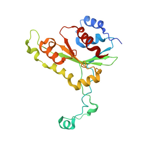
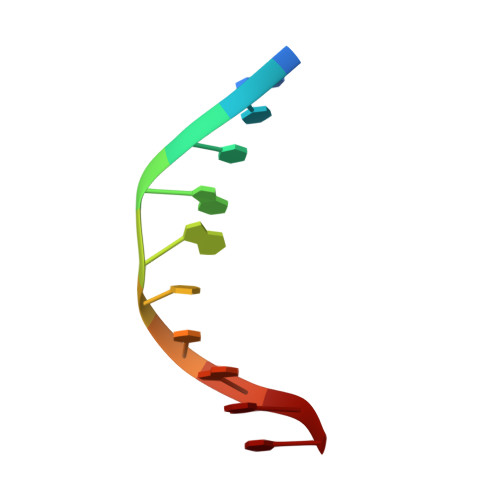
1D02

CRYSTAL STRUCTURE OF MUNI RESTRICTION ENDONUCLEASE IN COMPLEX WITH COGNATE DNA


Experimental Data Snapshot

  • Method: X-RAY DIFFRACTION
  • Resolution: 1.70 Å
  • R-Value Free: 0.213 
  • R-Value Work: 0.180 
  • R-Value Observed: 0.180 

wwPDB Validation   3D Report Full Report


This is version 1.4 of the entry. See complete history


Literature

Crystal structure of MunI restriction endonuclease in complex with cognate DNA at 1.7 A resolution.

Deibert, M.Grazulis, S.Janulaitis, A.Siksnys, V.Huber, R.

(1999) EMBO J 18: 5805-5816

  • DOI: https://doi.org/10.1093/emboj/18.21.5805
  • Primary Citation of Related Structures:  
    1D02

  • PubMed Abstract: 

    The MunI restriction enzyme recognizes the palindromic hexanucleotide sequence C/AATTG (the '/' indicates the cleavage site). The crystal structure of its active site mutant D83A bound to cognate DNA has been determined at 1.7 A resolution. Base-specific contacts between MunI and DNA occur exclusively in the major groove. While DNA-binding sites of most other restriction enzymes are comprised of discontinuous sequence segments, MunI combines all residues involved in the base-specific contacts within one short stretch (residues R115-R121) located at the N-terminal region of the 3(10)4 helix. The outer CG base pair of the recognition sequence is recognized solely by R115 through hydrogen bonds made by backbone and side chain atoms to both bases. The mechanism of recognition of the central AATT nucleotides by MunI is similar to that of EcoRI, which recognizes the G/AATTC sequence. The local conformation of AATT deviates from the typical B-DNA form and is remarkably similar to EcoRI-DNA. It appears to be essential for specific hydrogen bonding and recognition by MunI and EcoRI.


  • Organizational Affiliation

    Max-Planck-Institut für Biochemie, D-82152 Planegg-Martinsried, Germany. deibert@biochem.mpg.de


Macromolecules

Find similar proteins by:  (by identity cutoff)  |  3D Structure
Entity ID: 2
MoleculeChains Sequence LengthOrganismDetailsImage
TYPE II RESTRICTION ENZYME MUNIC [auth A],
D [auth B]
202MycoplasmaMutation(s): 1 
EC: 3.1.21.4
UniProt
Find proteins for P43642 (Mycoplasma sp)
Explore P43642 
Go to UniProtKB:  P43642
Entity Groups  
Sequence Clusters30% Identity50% Identity70% Identity90% Identity95% Identity100% Identity
UniProt GroupP43642
Sequence Annotations
Expand
  • Reference Sequence

Find similar nucleic acids by:  Sequence   |   3D Structure  

Entity ID: 1
MoleculeChains LengthOrganismImage
DNA (5'-D(*GP*CP*CP*AP*AP*TP*TP*GP*GP*C)-3')A [auth C],
B [auth D]
10N/A
Sequence Annotations
Expand
  • Reference Sequence
Experimental Data & Validation

Experimental Data

  • Method: X-RAY DIFFRACTION
  • Resolution: 1.70 Å
  • R-Value Free: 0.213 
  • R-Value Work: 0.180 
  • R-Value Observed: 0.180 
  • Space Group: P 21 21 21
Unit Cell:
Length ( Å )Angle ( ˚ )
a = 58.4α = 90
b = 72.47β = 90
c = 121.67γ = 90
Software Package:
Software NamePurpose
MLPHAREphasing
CNSrefinement
MOSFLMdata reduction
CCP4data scaling

Structure Validation

View Full Validation Report


Entry History 

Deposition Data

Revision History  (Full details and data files)

  • Version 1.0: 2000-03-08
    Type: Initial release
  • Version 1.1: 2008-04-27
    Changes: Version format compliance
  • Version 1.2: 2011-07-13
    Changes: Version format compliance
  • Version 1.3: 2018-03-14
    Changes: Database references
  • Version 1.4: 2024-02-07
    Changes: Data collection, Database references