Crystal structure of the hydrogenase maturating endopeptidase HYBD from Escherichia coli.
Fritsche, E., Paschos, A., Beisel, H.G., Bock, A., Huber, R.(1999) J Mol Biol 288: 989-998
- PubMed: 10331925
- DOI: https://doi.org/10.1006/jmbi.1999.2719
- Primary Citation of Related Structures:
1CFZ - PubMed Abstract:
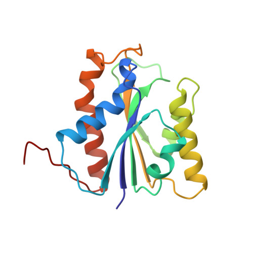
The maturation of [NiFe] hydrogenases includes formation of the nickel metallocenter, proteolytic processing of the metal center carrying large subunit, and its assembling with other hydrogenase subunits. The hydrogenase maturating enzyme HYBD from Escherichia coli, a protease of molecular mass 17.5 kDa, specifically cleaves off a 15 amino acid peptide from the C terminus of the precursor of the large subunit of hydrogenase 2 in a nickel-dependent manner. Here we report the crystal structure of HYBD at 2.2 A resolution. It consists of a twisted five-stranded beta-sheet surrounded by four and three helices, respectively, on each side. A cadmium ion from the crystallization buffer binds to the proposed nickel-binding site and is penta-coordinated by Glu16, Asp62, His93, and a water molecule in a pseudo-tetragonal arrangement. HYBD is topologically related to members of the metzincins superfamily of zinc endoproteinases, sharing the central beta-sheet and three helices. In contrast to the metzincins, the metal-binding site of HYBD is localized at the C-terminal end of the beta-sheet. Three helical insertions unique to HYBD pack against one side of the sheet, build up the active site cleft, and provide His93 as ligand to the metal. From this structure, we derive molecular clues into how the protease HYBD is involved in the hydrogenase maturation process.
Organizational Affiliation:
Max-Planck-Institut für Biochemia, Abteilung für Strukturforschung, Martinsried, D-82152, Germany. fritche@biochem.mpg.de