1CEL

THE THREE-DIMENSIONAL CRYSTAL STRUCTURE OF THE CATALYTIC CORE OF CELLOBIOHYDROLASE I FROM TRICHODERMA REESEI


Experimental Data Snapshot

  • Method: X-RAY DIFFRACTION
  • Resolution: 1.80 Å
  • R-Value Work: 0.181 
  • R-Value Observed: 0.181 

wwPDB Validation   3D Report Full Report


This is version 2.1 of the entry. See complete history


Literature

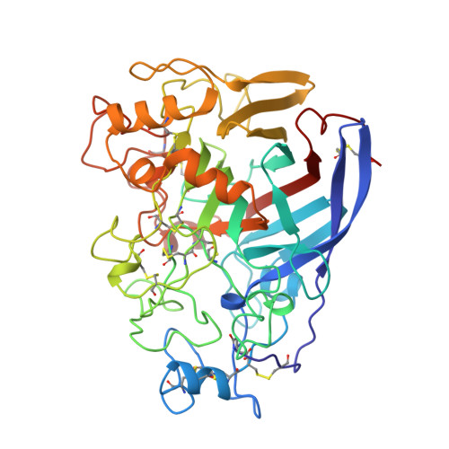
The three-dimensional crystal structure of the catalytic core of cellobiohydrolase I from Trichoderma reesei.

Divne, C.Stahlberg, J.Reinikainen, T.Ruohonen, L.Pettersson, G.Knowles, J.K.Teeri, T.T.Jones, T.A.

(1994) Science 265: 524-528

  • DOI: https://doi.org/10.1126/science.8036495
  • Primary Citation of Related Structures:  
    1CEL

  • PubMed Abstract: 

    Cellulose is the major polysaccharide of plants where it plays a predominantly structural role. A variety of highly specialized microorganisms have evolved to produce enzymes that either synergistically or in complexes can carry out the complete hydrolysis of cellulose. The structure of the major cellobiohydrolase, CBHI, of the potent cellulolytic fungus Trichoderma reesei has been determined and refined to 1.8 angstrom resolution. The molecule contains a 40 angstrom long active site tunnel that may account for many of the previously poorly understood macroscopic properties of the enzyme and its interaction with solid cellulose. The active site residues were identified by solving the structure of the enzyme complexed with an oligosaccharide, o-iodobenzyl-1-thio-beta-cellobioside. The three-dimensional structure is very similar to a family of bacterial beta-glucanases with the main-chain topology of the plant legume lectins.


  • Organizational Affiliation

    Department of Molecular Biology, Uppsala University, Sweden.


Macromolecules
Find similar proteins by:  (by identity cutoff)  |  3D Structure
Entity ID: 1
MoleculeChains Sequence LengthOrganismDetailsImage
1,4-BETA-D-GLUCAN CELLOBIOHYDROLASE I
A, B
434Trichoderma reeseiMutation(s): 0 
EC: 3.2.1.91
UniProt
Find proteins for P62694 (Hypocrea jecorina)
Explore P62694 
Go to UniProtKB:  P62694
Entity Groups  
Sequence Clusters30% Identity50% Identity70% Identity90% Identity95% Identity100% Identity
UniProt GroupP62694
Sequence Annotations
Expand
  • Reference Sequence
Small Molecules
Modified Residues  1 Unique
IDChains TypeFormula2D DiagramParent
PCA
Query on PCA
A, B
L-PEPTIDE LINKINGC5 H7 N O3GLN
Experimental Data & Validation

Experimental Data

  • Method: X-RAY DIFFRACTION
  • Resolution: 1.80 Å
  • R-Value Work: 0.181 
  • R-Value Observed: 0.181 
  • Space Group: P 21 21 2
Unit Cell:
Length ( Å )Angle ( ˚ )
a = 84α = 90
b = 86.2β = 90
c = 111.8γ = 90
Software Package:
Software NamePurpose
X-PLORmodel building
X-PLORrefinement
X-PLORphasing

Structure Validation

View Full Validation Report


Entry History 

Deposition Data

Revision History  (Full details and data files)

  • Version 1.0: 1994-11-01
    Type: Initial release
  • Version 1.1: 2008-03-10
    Changes: Version format compliance
  • Version 1.2: 2011-07-13
    Changes: Non-polymer description, Version format compliance
  • Version 1.3: 2017-11-29
    Changes: Derived calculations, Other
  • Version 2.0: 2019-12-25
    Changes: Data collection, Derived calculations, Polymer sequence
  • Version 2.1: 2020-07-29
    Type: Remediation
    Reason: Carbohydrate remediation
    Changes: Advisory, Data collection, Derived calculations, Structure summary