1B7E

TRANSPOSASE INHIBITOR


Experimental Data Snapshot

  • Method: X-RAY DIFFRACTION
  • Resolution: 2.90 Å
  • R-Value Work: 0.195 

wwPDB Validation   3D Report Full Report


This is version 1.4 of the entry. See complete history


Literature

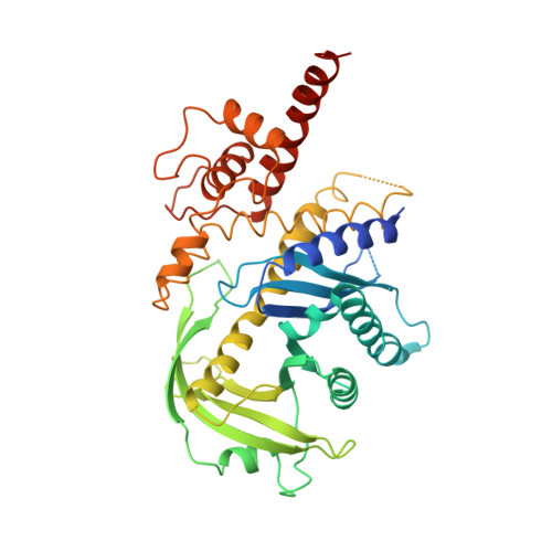
The three-dimensional structure of a Tn5 transposase-related protein determined to 2.9A resolution.

Davies, D.R.Braam, L.M.Reznikoff, W.S.Rayment, I.

(1999) J Biol Chem 274: 11904-11913

  • DOI: https://doi.org/10.1074/jbc.274.17.11904
  • Primary Citation of Related Structures:  
    1B7E

  • PubMed Abstract: 

    Transposon Tn5 employs a unique means of self-regulation by expressing a truncated version of the transposase enzyme that acts as an inhibitor. The inhibitor protein differs from the full-length transposase only by the absence of the first 55 N-terminal amino acid residues. It contains the catalytic active site of transposase and a C-terminal domain involved in protein-protein interactions. The three-dimensional structure of Tn5 inhibitor determined to 2.9-A resolution is reported here. A portion of the protein fold of the catalytic core domain is similar to the folds of human immunodeficiency virus-1 integrase, avian sarcoma virus integrase, and bacteriophage Mu transposase. The Tn5 inhibitor contains an insertion that extends the beta-sheet of the catalytic core from 5 to 9 strands. All three of the conserved residues that make up the "DDE" motif of the active site are visible in the structure. An arginine residue that is strictly conserved among the IS4 family of bacterial transposases is present at the center of the active site, suggesting a catalytic motif of "DDRE." A novel C-terminal domain forms a dimer interface across a crystallographic 2-fold axis. Although this dimer represents the structure of the inhibited complex, it provides insight into the structure of the synaptic complex.


  • Organizational Affiliation

    Institute for Enzyme Research and Department of Biochemistry, University of Wisconsin, Madison, Wisconsin 53705, USA.


Macromolecules
Find similar proteins by:  (by identity cutoff)  |  3D Structure
Entity ID: 1
MoleculeChains Sequence LengthOrganismDetailsImage
PROTEIN (TRANSPOSASE INHIBITOR PROTEIN FROM TN5)420Escherichia coliMutation(s): 0 
UniProt
Find proteins for Q46731 (Escherichia coli)
Explore Q46731 
Go to UniProtKB:  Q46731
Entity Groups  
Sequence Clusters30% Identity50% Identity70% Identity90% Identity95% Identity100% Identity
UniProt GroupQ46731
Sequence Annotations
Expand
  • Reference Sequence
Small Molecules
Ligands 1 Unique
IDChains Name / Formula / InChI Key2D Diagram3D Interactions
TPT
Query on TPT

Download Ideal Coordinates CCD File 
B [auth A],
C [auth A]
2,2':6',2''-TERPYRIDINE PLATINUM(II) Chloride
C15 H11 Cl N3 Pt
XTQMQLRZXBEQCS-UHFFFAOYSA-M
Experimental Data & Validation

Experimental Data

  • Method: X-RAY DIFFRACTION
  • Resolution: 2.90 Å
  • R-Value Work: 0.195 
  • Space Group: P 21 21 2
Unit Cell:
Length ( Å )Angle ( ˚ )
a = 181.8α = 90
b = 71.9β = 90
c = 41.3γ = 90
Software Package:
Software NamePurpose
DENZOdata reduction
SCALEPACKdata scaling
SOLVEphasing
TNTrefinement
HKL-2000data reduction

Structure Validation

View Full Validation Report


Entry History 

Deposition Data

Revision History  (Full details and data files)

  • Version 1.0: 1999-04-21
    Type: Initial release
  • Version 1.1: 2007-10-16
    Changes: Version format compliance
  • Version 1.2: 2011-07-13
    Changes: Version format compliance
  • Version 1.3: 2017-10-04
    Changes: Refinement description
  • Version 1.4: 2022-12-21
    Changes: Database references, Derived calculations