1B6U

CRYSTAL STRUCTURE OF THE HUMAN KILLER CELL INHIBITORY RECEPTOR (KIR2DL3) SPECIFIC FOR HLA-CW3 RELATED ALLELES


Experimental Data Snapshot

  • Method: X-RAY DIFFRACTION
  • Resolution: 3.00 Å
  • R-Value Free: 0.320 
  • R-Value Work: 0.248 
  • R-Value Observed: 0.248 

wwPDB Validation   3D Report Full Report


This is version 1.3 of the entry. See complete history


Literature

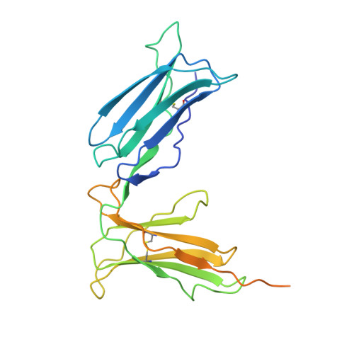
Crystal structure of the human p58 killer cell inhibitory receptor (KIR2DL3) specific for HLA-Cw3-related MHC class I.

Maenaka, K.Juji, T.Stuart, D.I.Jones, E.Y.

(1999) Structure 7: 391-398

  • DOI: https://doi.org/10.1016/s0969-2126(99)80052-5
  • Primary Citation of Related Structures:  
    1B6U

  • PubMed Abstract: 

    T cells and natural killer (NK) cells perform complementary roles in the cellular immune system. T cells identify infected cells directly through recognition of antigenic peptides that are displayed at the target cell surface by the classical major histocompatibility complex (MHC) class I molecules. NK cells monitor the target cell surface for malfunction of this display system, lysing potentially infected cells that might otherwise evade recognition by the T cells. Human killer cell inhibitory receptors (KIRs) control this process by either inhibiting or activating the cytotoxic activity of NK cells via specific binding to MHC class I molecules on the target cell.


  • Organizational Affiliation

    Laboratory of Molecular Biophysics, Rex Richards Building, South Parks Road, Oxford OX1 3QU, UK. katsumi@biop.ox.ac.uk


Macromolecules
Find similar proteins by:  (by identity cutoff)  |  3D Structure
Entity ID: 1
MoleculeChains Sequence LengthOrganismDetailsImage
P58 KILLER CELL INHIBITORY RECEPTOR257Homo sapiensMutation(s): 0 
Gene Names: NKAT2
UniProt & NIH Common Fund Data Resources
Find proteins for P43628 (Homo sapiens)
Explore P43628 
Go to UniProtKB:  P43628
PHAROS:  P43628
GTEx:  ENSG00000243772 
Entity Groups  
Sequence Clusters30% Identity50% Identity70% Identity90% Identity95% Identity100% Identity
UniProt GroupP43628
Sequence Annotations
Expand
  • Reference Sequence
Experimental Data & Validation

Experimental Data

  • Method: X-RAY DIFFRACTION
  • Resolution: 3.00 Å
  • R-Value Free: 0.320 
  • R-Value Work: 0.248 
  • R-Value Observed: 0.248 
  • Space Group: P 65 2 2
Unit Cell:
Length ( Å )Angle ( ˚ )
a = 95.4α = 90
b = 95.4β = 90
c = 130.8γ = 120
Software Package:
Software NamePurpose
DENZOdata reduction
SCALEPACKdata scaling
CNSrefinement
CNSphasing

Structure Validation

View Full Validation Report


Entry History 

Deposition Data

Revision History  (Full details and data files)

  • Version 1.0: 1999-01-27
    Type: Initial release
  • Version 1.1: 2008-03-24
    Changes: Version format compliance
  • Version 1.2: 2011-07-13
    Changes: Version format compliance
  • Version 1.3: 2018-06-13
    Changes: Data collection