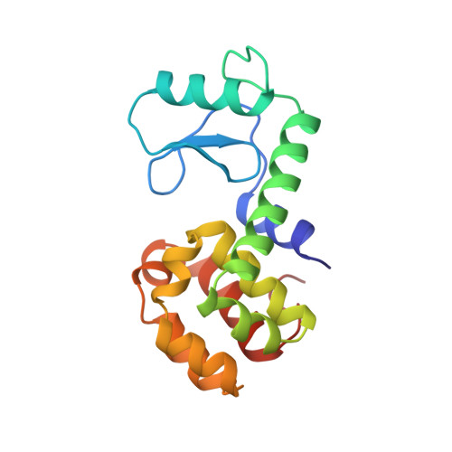
Specificity of ligand binding in a buried nonpolar cavity of T4 lysozyme: linkage of dynamics and structural plasticity.
Morton, A., Matthews, B.W.(1995) Biochemistry 34: 8576-8588
- PubMed: 7612599
- DOI: https://doi.org/10.1021/bi00027a007
- Primary Citation of Related Structures:
181L, 182L, 183L, 184L, 185L, 186L, 187L, 188L, 1NHB - PubMed Abstract:
To better understand the role of shape complementarity in ligand binding and protein core interactions, the structures have been determined of a set of ligands bound within a cavity-containing mutant of T4 lysozyme. The interior cavity is seen to consist of two parts that respond very differently to the binding of ligands. First, there is a relatively rigid region that does not relax significantly upon binding any ligand. Second, there is a more flexible region that moves to various extents in response to binding the different ligands. The part of the binding site that remains rigid is characterized by low temperature factors and strong protection from hydrogen exchange. This part of the site appears to be primarily responsible for discriminating between ligands of different shape (i.e., for determining specificity). The more flexible region, characterized by relatively high temperature factors and weak protection from hydrogen exchange, allows some promiscuity in binding by undergoing variable amounts of deformation at essentially the same energetic cost. This linkage between the dynamic information represented by crystallographic temperature factors and hydrogen-exchange behavior on the one hand, and structural plasticity in response to ligand binding on the other hand, suggests a way to improve our understanding of steric interactions in protein cores and protein-ligand binding sites. Ligand design and packing algorithms might take advantage of this information, requiring complementary interactions where the protein is rigid and allowing some overlap in regions where the protein is flexible.
Organizational Affiliation:
Institute of Molecular Biology, Howard Hughes Medical Institute, Eugene, Oregon, USA.