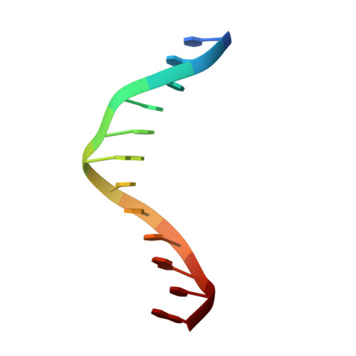
Crystal and molecular structure of d(CGTAGATCTACG) at 2.25 A resolution.
Leonard, G.A., Hunter, W.N.(1993) J Mol Biol 234: 198-208
- PubMed: 8230199
- DOI: https://doi.org/10.1006/jmbi.1993.1574
- Primary Citation of Related Structures:
119D - PubMed Abstract:
Single crystal X-ray diffraction methods have been used to determine the structure of the synthetic DNA dodecamer d(CGTAGATCTACG). The dodecamer presents a new crystal form of B-DNA in the monoclinic system, space group C2 with a = 64.83 A, b = 35.36 A, c = 25.35 A and beta = 92.24 degrees. Structure solution was by molecular replacement and the refinement used a combination of rigid body treatment, molecular dynamics simulated annealing and restrained least-squares methods. The refinement has been concluded with an R-factor of 13.8% for 2120 reflections (78% of what is theoretically available) with F > or = 2 sigma (F) in the resolution range 7.0 to 2.25 A. The asymmetric unit comprises a B-form duplex (24 nucleotides, molecular weight 7.2 kDa), 136.5 water molecules and a single magnesium ion at 50% occupancy. The DNA model has root-mean-square derivations from standard bond lengths of 0.011 A. The central part of the structure is d(GATC), a tetrad sequence involved in methylation and mismatch repair. A comparison with other structures containing such a tetrad indicates polymorphism at the tetrad level. In our structure a narrow minor groove with a pronounced hydration pattern is observed. Such features may be of importance in the recognition of the sequence by specific enzymes. A noteworthy feature of the hydration is the identification of several pentagonal arrangements of hydrogen bonding groups. Details of conformational parameters, base stacking patterns, intermolecular interactions and hydration are presented and comparisons with related structures are given.
Organizational Affiliation:
Department of Chemistry, University of Manchester, U.K.